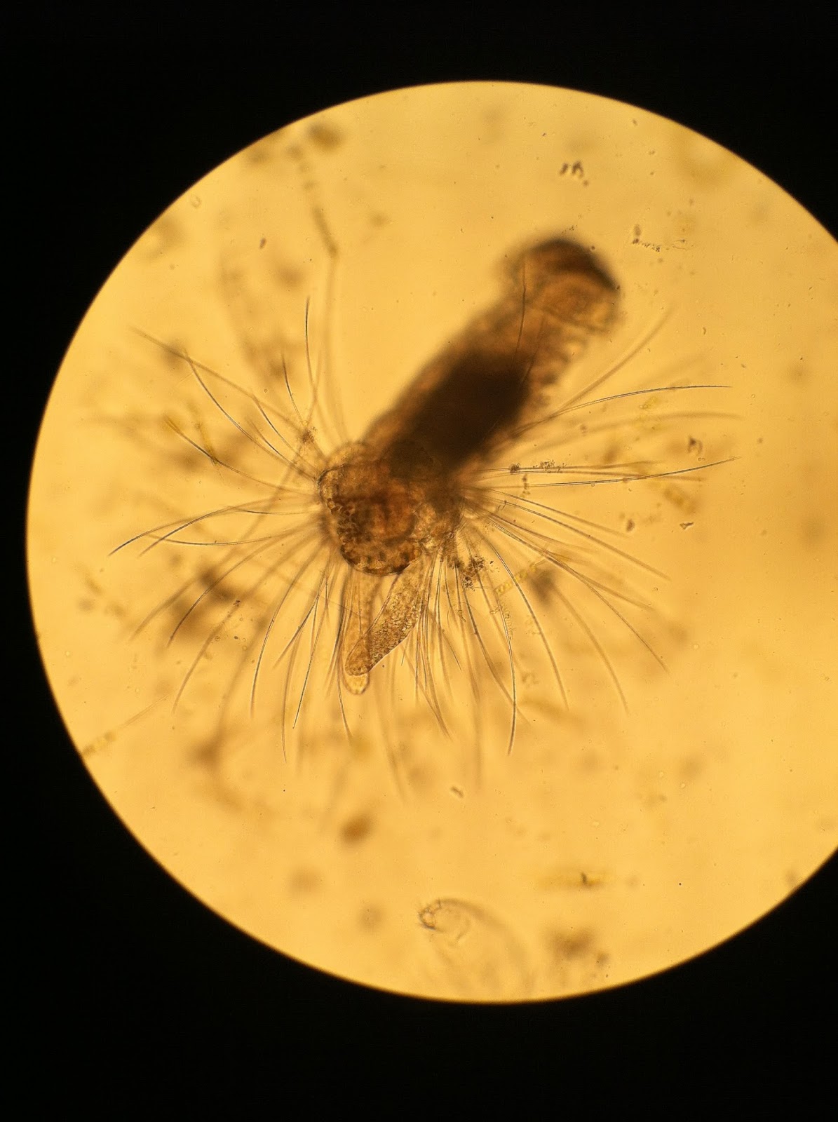
A lot of my time has been spent learning to identify plankton through the microscopes. I've found I can get somewhat decent photos of what I'm seeing by holding my phone's camera up to the eyepiece. I'm posting these because I think the intricate forms are amazing, and to share what I've been seeing.
First, the phytoplankton (plants):
 |
| Protoperidinium |
.jpg) |
| Asterionellopsis |
 |
| Coscinodiscus |
.JPG) |
| Ditylum |
.jpg) |
| Dictyocha |
 |
| Eucampia, Pseudonitzschia, Chaetoceros, Coscinodiscus, Probiscia |
 |
| Skeletonema |
 |
| Stephanopyxis |
 |
| Odontella |
 |
| Thalasionema (center) |
Next, the zooplankton (animals):
.JPG) |
| Adult Copepod, side view |
 |
| Adult Copepod, top view |
 |
| Copepod Nauplius |
 |
| Cypris Larva |
 |
| Rotifer |
 |
| Another Rotifer |
 |
| Shrimp Larva |
 |
| Polychaete |
 |
| Barnacle Nauplius |
 |
| Brachiolaria (second larval stage of sea star) amidst a lot of phytoplankton |
 |
| Brachiolaria |
 |
| Tintinnid |
And, views of both zooplankton and phytoplankton from both dissecting scope and compound scope:
 |
| Ctenophore |
 |
| A ctenophore's pharynx, stomach, and tentacle bases |
 |
| A ctenophore's retracted tentacle |
Finally, a few movies through the microscope:
Tracking a Dinoflagellate
The Undulations of a Polychaete
The Brachiolaria
The Living Sea, in the compound microscope
The Living Sea, in the dissecting microscope

.jpg)

.JPG)
.jpg)


.JPG)


Awesome pictures Peter!
ReplyDeleteHi, thank you so much for sharing these images. They are incredible. Do you ever use stains to better visualize the critters?
ReplyDelete