The past week in the Maslakova lab has been full of new discoveries and exciting plans. By far the most interesting development has been the identification of an obvious phenotype (a physical characteristic) of pilidium larvae lacking a very specific transcription factor. These larvae were depleted of this protein by injecting a number of zygotes (fertilized eggs) with a morpholino designed to eliminate the production of this protein specifically.
First a little background about what this protein does, and why we care:
First a little background about what this protein does, and why we care:
| A transcription factor binding to DNA |
 |
| Pilidium larva with juvenile worm |
However, transcription factors are fairly ubiquitous within the genome of any given organism - so why this specific one? Simply put - this gene had already been cloned in M. alaskensis by Ph.D. student Laurel Hiebert and this made it very easy to order a morpholino to knock it out. Furthermore, morpholino knockouts have never been tried before in M. alaskensis, and oftentimes embryos can be extremely resistant to morpholino treatment. This transcription factor seemed as good a place to start as any, considering that we were not 100% sure that the morpholinos would even work. However, morpholino treatment seems to be working extremely effectively and we may even be able to target other developmental pathways before the end of the summer.
This coming week will be largely spent addressing the phenotype identified in the larvae that we have depleted of this protein. I will likely be using more specific staining techniques and confocal microscopy to determine exactly what is going on in depleted larvae, compared to normal healthy larvae. We will also be continuing to address the phenotype identified in MKLP1 depleted larvae. As I mentioned in a previous blog post, MKLP1 is a motor protein crucial for a cell to identify its mid-line and undergo cell division. We will also be using fluorescent markers to identify any irregularities in these cells before they lose the ability to undergo cell division - these irregularities will provide clues as to what this protein does and how it interacts with other players in the cell division process.
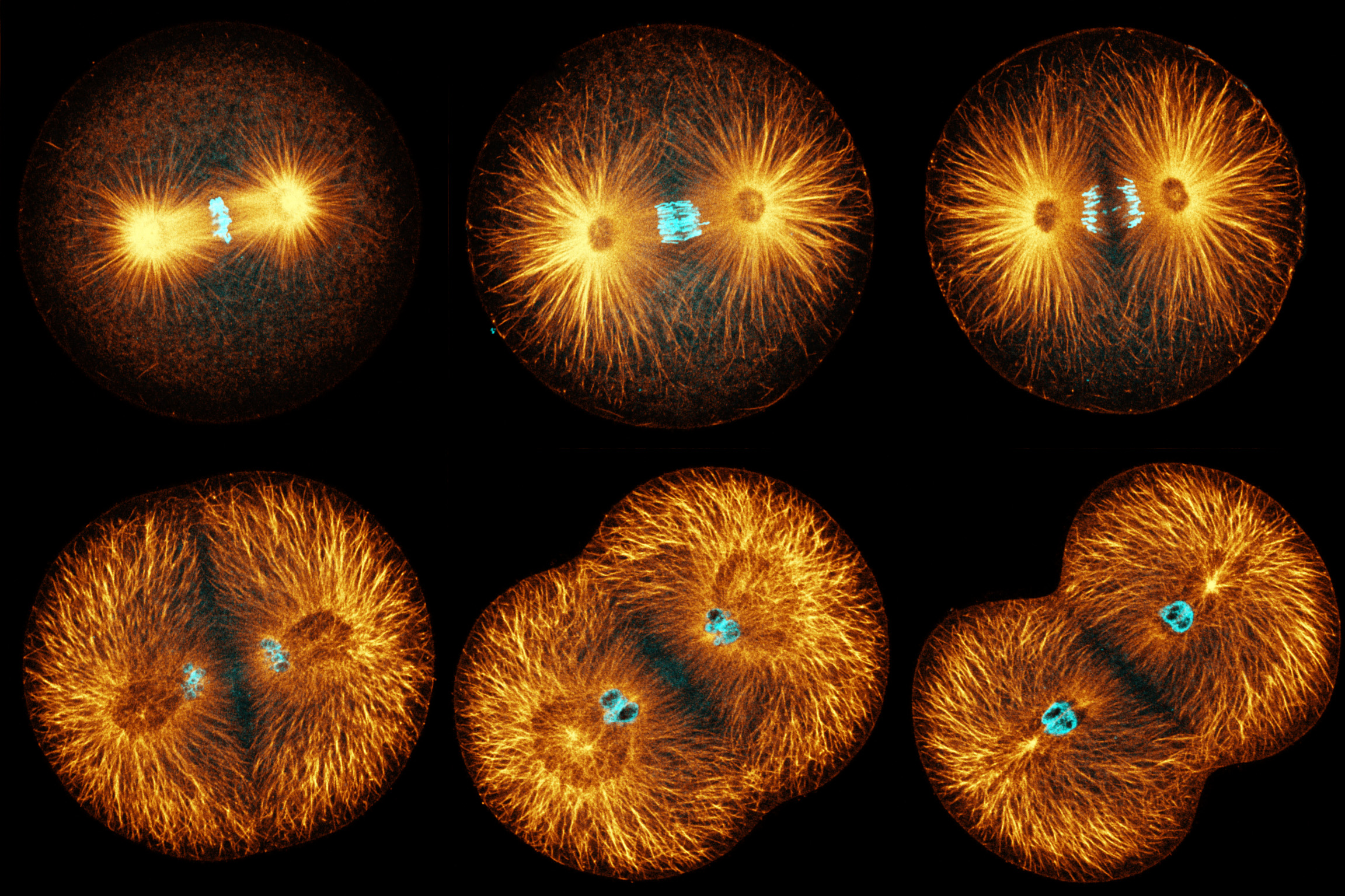 |
| Time-lapse sequence of cell division - photo by George von Dassow, senior research associate at the OIMB |
More results to come soon!
Excellent - I'm glad the oligos are working for you!
ReplyDelete- Jon at Gene Tools
Wow what an interesting project. Great discussion of transcription factors and thanks for the pictures of mitosis!
ReplyDelete